JavaScript is disabled for your browser. Some features of this site may not work without it.
- ResearchSpace
- →
- Research Publications/Outputs
- →
- Conference Publications
- →
- View Item
| dc.contributor.author |
Karsten, AE

|
|
| dc.contributor.author |
Singh, A
|
|
| dc.contributor.author |
Ndhundhuma, I
|
|
| dc.date.accessioned | 2010-07-16T13:29:51Z | |
| dc.date.available | 2010-07-16T13:29:51Z | |
| dc.date.issued | 2010-01 | |
| dc.identifier.citation | Karsten, AE, Singh, A, and Ndhundhuma, I. 2010. Optical coherence tomography as a research tool for biomaterials. European Cells and Materials, Vol. 19(Suppl. 1), pp 18 | en |
| dc.identifier.issn | 1473-2262 | |
| dc.identifier.uri | http://www.ecmjournal.org/journal/supplements/vol019supp01/pdf/v019supp01a018.pdf | |
| dc.identifier.uri | http://hdl.handle.net/10204/4100 | |
| dc.description | Copyright: 2010 The authors | en |
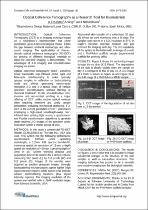
| dc.description.abstract | In this study a commercial FD-OCT (Model OCM1300SS) by Thorlabs Inc., USA was used. This system has the following performance specifications: 1300nm centre operating wavelength, a 110nm 3dB spectral bandwidth, transverse spatial air resolution of 15-um, a depth spatial air resolution of 20 um, a probing depth of 3mm in air, 25mm working distance and acquisition time of 30 seconds for a sample volume measuring 512 pixels (L) by 512 pixels (W) and 512 pixels (D). Images of the samples were acquired as positive grayscale images. Strongly backscattering microstructures appear lighter (high signal intensity regions) while darker areas indicate weaker backscattering locations (low signal intensity regions). The imaging resolution of the system was verified with a USAF resolution target from Edmund Scientific, USA. Abdominal skin samples of a euthanized, 32 days old Wistar rat were monitored over a 15 days. The samples were stored in a CO2 incubator at 37°C in Eagle’s minimal essential medium and just removed for imaging each day. The 3-D capability of the system is illustrated with an image of a moth and a PNIPAAm scaffold sample (experimental scaffold for growing cells). | en |
| dc.language.iso | en | en |
| dc.publisher | The authors | en |
| dc.subject | Optical coherence tomography | en |
| dc.subject | Biomaterials | en |
| dc.subject | Volume measuring pixels | en |
| dc.subject | High signal intensity | en |
| dc.subject | Low signal intensity | en |
| dc.title | Optical coherence tomography as a research tool for biomaterials | en |
| dc.type | Conference Presentation | en |
| dc.identifier.apacitation | Karsten, A., Singh, A., & Ndhundhuma, I. (2010). Optical coherence tomography as a research tool for biomaterials. The authors. http://hdl.handle.net/10204/4100 | en_ZA |
| dc.identifier.chicagocitation | Karsten, AE, A Singh, and I Ndhundhuma. "Optical coherence tomography as a research tool for biomaterials." (2010): http://hdl.handle.net/10204/4100 | en_ZA |
| dc.identifier.vancouvercitation | Karsten A, Singh A, Ndhundhuma I, Optical coherence tomography as a research tool for biomaterials; The authors; 2010. http://hdl.handle.net/10204/4100 . | en_ZA |
| dc.identifier.ris | TY - Conference Presentation AU - Karsten, AE AU - Singh, A AU - Ndhundhuma, I AB - In this study a commercial FD-OCT (Model OCM1300SS) by Thorlabs Inc., USA was used. This system has the following performance specifications: 1300nm centre operating wavelength, a 110nm 3dB spectral bandwidth, transverse spatial air resolution of 15-um, a depth spatial air resolution of 20 um, a probing depth of 3mm in air, 25mm working distance and acquisition time of 30 seconds for a sample volume measuring 512 pixels (L) by 512 pixels (W) and 512 pixels (D). Images of the samples were acquired as positive grayscale images. Strongly backscattering microstructures appear lighter (high signal intensity regions) while darker areas indicate weaker backscattering locations (low signal intensity regions). The imaging resolution of the system was verified with a USAF resolution target from Edmund Scientific, USA. Abdominal skin samples of a euthanized, 32 days old Wistar rat were monitored over a 15 days. The samples were stored in a CO2 incubator at 37°C in Eagle’s minimal essential medium and just removed for imaging each day. The 3-D capability of the system is illustrated with an image of a moth and a PNIPAAm scaffold sample (experimental scaffold for growing cells). DA - 2010-01 DB - ResearchSpace DP - CSIR KW - Optical coherence tomography KW - Biomaterials KW - Volume measuring pixels KW - High signal intensity KW - Low signal intensity LK - https://researchspace.csir.co.za PY - 2010 SM - 1473-2262 T1 - Optical coherence tomography as a research tool for biomaterials TI - Optical coherence tomography as a research tool for biomaterials UR - http://hdl.handle.net/10204/4100 ER - | en_ZA |